Figure 1

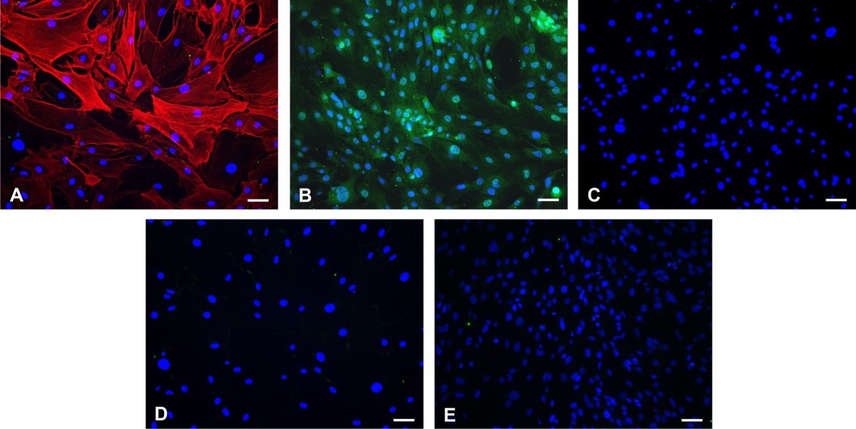
Determination of the purity of the pericyte culture. A primary culture of pericytes isolated from mouse brain microvessels was labeled with anti-α smooth muscle actin antibody (pericyte marker; red) (Panel A), anti-CD13 antibody (pericyte marker; green) (Panel B), anti-GFAP antibody (astrocytes marker; green) (Panel C), Griffonia simplicifolia lectin (microglial marker; green) (Panel D) or anti-factor VIII antibody (endothelial cell marker; green) (Panel E) and counterstained with nuclear stain DAPI (blue). Visual observation of immunostained cells in pericyte cultures demonstrates that they primarily consist of a α-smooth muscle actin/CD13 positive pericytes. No contamination with microglia, astrocytes or endothelial cells was detected. Scale bar: 40 μm.
