Fig. 2

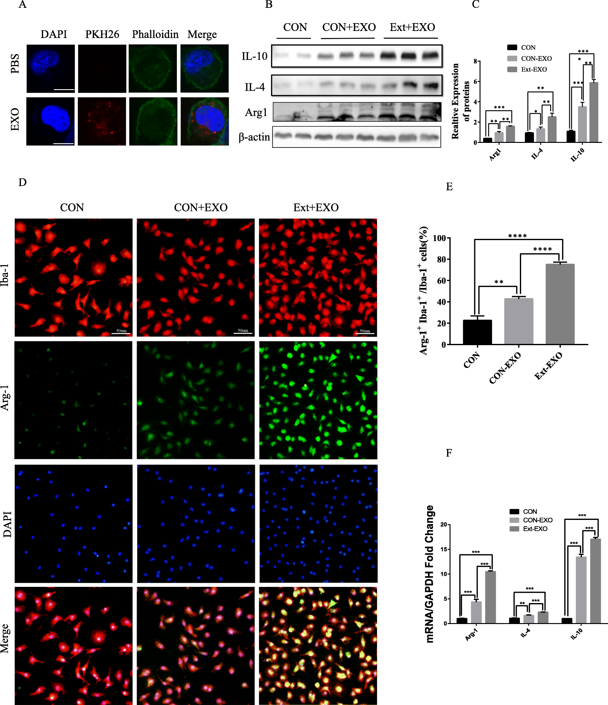
Astrocyte-derived exosomes are taken up by primary microglia and promote microglial M2 phenotype transformation. a PKH26 staining and exosome uptake. It was shown by confocal microscopy that exosomes (red) were taken up by the cells and existed in the cytoplasm and around the cell nucleus. Immunofluorescence of the primary microglia showing DAPI (blue), exosomes (red) and F-actin (green). No red fluorescent signal was detected in the PBS control group. PBS: microglial staining with PBS. Exo: the exosomes labelled with pkh26 were incubated with microglia for 24 h. Bar = 10 μm. b, c Primary cultured microglia were divided into three groups: con, con+Exo and Ext+Exo. Exo: The microglia were incubated with astrocyte-derived exosomes for 4 h. Ext+Exo: primary cultured microglia were stimulated with “brain extracts” for 24 h and washed before incubation with exosomes. These M2 markers were also detected by Western blot analysis. d, e Fluorescence confocal microscopic images showing both Iba-1 (microglia marker) and Arg-1 (M2 marker) expression increased after astrocyte-derived exosome treatment. Bar = 50 μm. Quantification of the percentage of Arg-1+Iba-1+ cells among total Iba-1+ cells. This effect was more significant after “brain extract” stimulation. f The mRNA expression of microglia M2 markers (Arg-1, IL-4, IL-10) was detected by RT-PCR (data were presented as mean ± SD, *p < 0.05, **p < 0.01, ***p < 0.001, n = 5, one-way ANOVA)
