Figure 4
From: Plasminogen in cerebrospinal fluid originates from circulating blood

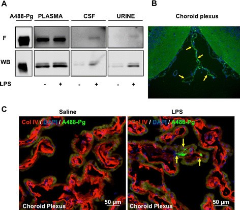
In vivo evidence of the circulating origin of plasminogen in cerebrospinal fluid. Samples were obtained from rats injected with plasminogen labelled with Alexa Fluor 488 dye (A488-Pg). Group L2 rats were challenged with lipopolysaccharide (LPS), and group C2 was challenged with saline (see flowchart in Figure 1). (A). An equal volume (10 μl) of either plasma diluted 1:50 or of cerebrospinal fluid (CSF) or urine was electrophoresed, and fluorescence (F) in the gel was directly revealed using ImageQuant TL 7.0 image analysis software (upper panel). The gel was then transferred onto a polyvinylidene fluoride membrane and detected by Western blotting with a rabbit antibody to mouse plasminogen (WB, lower panel). Representative samples are shown. (B) Micrograph showing the presence of circulating A488-Pg (indicated by arrows) in the choroid plexus of an LPS-treated rats detected by direct fluorescence microscopy. 4’,6-diamidino-2-phenylindole (DAPI) staining (blue) indicates cell nuclei. (C) Magnified images of choroid plexus of saline- and LPS-treated rats showing the presence of circulating A488-Pg (indicated by yellow arrows) only in the LPS condition, as detected by direct fluorescence microscopy. DAPI staining (blue) indicates cell nuclei, and collagen type IV (Col IV, red) is used as a vessel marker.
